


To date, science knows about 280 species of worms that can develop and live in the human body, parasitizing various organs and tissues. The incidence of human worm infection depends on the climatic and socio-economic conditions of certain areas (lagging countries, especially those located in tropical and subtropical zones, have much higher levels of parasitic infections than economically developed countries).
Methods of human infection with helminths
- Biohelminthiasis (infection of animals).
- Infectious helminthiasis (spread from person to person).
- Geohelminthiasis (diseases caused by parasites that carry out one of their life cycles on earth).
Factors influencing the manifestations of helminthiasis
- The way the parasite enters the body;
- The degree of adaptation of the helminth to the human body;
- Population density (number) of parasitic individuals;
- The habitat of the worm (tissue parasites live in the thickness of soft tissues and luminescence in the lumen of hollow organs). Some helminths with different phases have both luminal and tissue forms. The larval and developing stages of worms usually cause more pronounced pathological changes.
In the absence of re-infection, the number of adult parasites does not increase in the human body. This property significantly distinguishes helminthic invasions from diseases caused by bacteria, viruses, fungi, and protozoa.
Worms in humans: symptoms
Helminthiasis is a disease characterized by two stages of the process (acute, from two weeks to two months) and chronic (from several months to several years).
Symptoms of the acute phase of helminthiasis
The first signs of the disease may appear at different times (most often after 2-3 weeks, in case of ascariasis - after 2-3 days, and in case of filariasis the incubation period can last from 6 to 18 months).
The most common symptom in the acute stage of parasite invasion is an allergic reaction (antibodies are formed against antigens from migratory parasite larvae). People infected with worms often have itchy rashes on the skin, are prone to recurrence, regional lymph node enlargement, generalized or local edema, and muscle and joint pain. Migratory parasitic larvae can cause chest pain, coughing, suffocation attacks, stool disorders, nausea, and vomiting.
However, the acute phase of helminthiasis may be associated with more severe disorders (pneumonia, hepatitis, allergic myocarditis, hepatosplenomegaly (enlarged liver and spleen), meningoencephalitis).
The number of eosinophils in the blood increases (eosinophilia) and the normal quantitative ratio between protein fractions is disturbed (dysproteinemia).
Signs of chronic helminthiasis
The symptoms of the chronic phase depend directly on which organ the parasites "live in" and their size and number play an important role.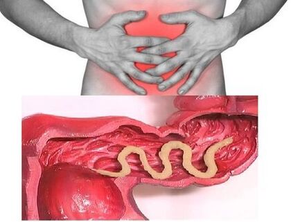 So, by parasitizing the intestines of individuals, the disease can be asymptomatic (except in cases infected with very large parasites). Dyspeptic disorders are characteristic signs of the chronic phase of intestinal helminthiasis. Asthenoneurotic and pain syndromes are more pronounced in children. With massive invasion of roundworms, intestinal obstruction, obstructive jaundice, and inflammation of the pancreas are possible.
Helminths cause indigestion and damage the absorption of vitamins, minerals, carbohydrates, proteins and fats by consuming all the substances needed for their vital activities from the host. At the same time, the waste products of worms inhibit the normal intestinal microflora and reduce the body's immune power.
People with helminthiasis have a significantly increased risk of malignancies due to a weakened immune system and an increased process of cell division (as a result of the constant repair of tissues damaged by the parasite).
Types of helminths parasitizing the human body
Human helminthiasis is caused by two types of worms: round (nematodes) and flat (tapeworms and spots).
Roundworms
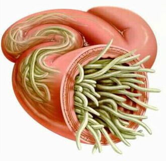
Pinworm
Enterobiasis parasites are small (up to 10 mm) thin hollow worms with an off-white color. The infection occurs through food (orally). This is due to dirty hands. The parasite's eggs may be in the ground, on the wool of infected animals, unwashed vegetables and fruits, and so on. However, in the case of enterobiasis, self-infection is common (especially in children), which is the ingestion of itchy areas and subsequent eggs.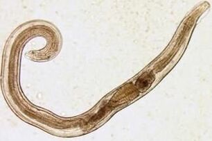 The pinworm larva develops in the digestive system within two weeks. As adults, the worm parasites the lower and upper sections of the colon.
Even at the larval stage, pinworm damages the body of its host, producing enzymes that irritate the intestinal walls and lead to an inflammatory process. Adult parasites adhere to or penetrate the deeper layers of the intestinal mucosa, disrupting its integrity and contributing to the conclusion of a secondary bacterial infection. Peritonitis may develop with pinworms perforation of the small intestinal wall. Irritation of intestinal receptors also disrupts the motor and secretory functions of the gastrointestinal tract, leading to gastroduodenitis, enteritis, and so on. It leads to its formation. In childhood, long-term enterobiasis can cause nerve disorders and delayed physical development.
Ascaris
Ascaris is a large, spindle-shaped parasite, red-yellow in color, reaching 40 cm (females) and 15-25 cm (males) in adulthood. Without suction cups or other fastening devices, the globular worm is able to move independently towards food masses. The eggs laid by the female of the parasite are excreted along with the feces.
Ascariasis infection occurs when ripe eggs are swallowed with water or unwashed vegetables and fruits with soil particles. Once the eggs enter the intestines, mature larvae emerge from them. They then penetrate the intestinal wall and travel through the bloodstream to the heart and from there to the lungs. Through the pulmonary alveoli, the cylindrical larva enters the oral cavity again through the airways. After repeated ingestion, the parasite reaches the small intestine where it develops into an adult. The worm lives for 12 months, then dies and is excreted along with the feces. One and hundreds of individuals can live in the gut of a farmer.
In the intestinal phase of their existence, spherical worms endowed with the ability of spiral movements can penetrate even the narrowest openings. This feature of the parasite often leads to the development of quite severe complications (obstructive jaundice or inflammation of the pancreas). Allergens secreted by globular worms can cause severe allergic reactions. A large number of adults can cause intestinal obstruction, and worms that enter the respiratory system sometimes cause suffocation.
Vlasoglav
Vlasoglav, the causative agent of trichocephalosis, is a white helminth 4-5 cm in size parasitizing in the early stages of the colon. The parasite feeds on the blood and tissues of the rectal mucosa.
Whipworm eggs laid by the female on the intestinal wall come out with the feces. Their development takes place in the environment (optimally in the soil). Eggs with the larvae of the parasite in them get into the body with food, dirty hands, water or unwashed vegetables and fruits.
Trichocephalosis is asymptomatic in a small number of worms. In the severe stage (with massive invasion), the patient causes abdominal pain, severe diarrhea, sometimes accompanied by rectal prolapse. This condition is most common in debilitated children. In the middle phase of trichocephalosis, growth retardation of the child is possible.
Trichinella
The causative agent of trichinosis is a small, round, 2-5 mm long helminth. Infection occurs with the consumption of poorly roasted meat (pork, bear meat, wild boar).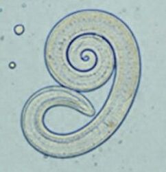 Penetrating into the intestines, the parasite larvae reach the state of a mature individual in 3-4 days. The worm has a lifespan of 40 days, after which the parasite dies. By piercing the intestinal wall, the larvae enter the bloodstream and reach all the organs of the human body, settling in the muscles. In this case, it most often affects the respiratory and facial muscles as well as the flexor muscles of the limbs.
In the first days after invasion, patients complain of abdominal pain. After about 2 weeks, the body temperature rises to 39-40 C, itchy rashes appear on the skin, muscle aches develop and the face swells. During this period, there is a significant risk of death in the event of a massive infection. After about a month, the patient recovers. The parasite is sealed in a spiral and then dies within two years.
Hookworm and nekator
These two parasites are similar in their biological properties as well as in the diseases they cause. In this respect, it is customary to combine them under a common name (hookworm). Worms 10-15 mm long parasitize in 12-p. gut. It should be noted that this is one of the most common, but at the same time quite rare parasites. Worm larvae enter the human body through the skin in contact with contaminated soil. Furthermore, once in the bloodstream, they migrate to the lungs like globules and then through the bronchi to the digestive system in addition to the sputum. An ankylostoma parasites the intestine and attaches to the intestinal wall. The blood-only parasite bites through the blood vessels that penetrate the mucosa, injecting an anticoagulant component there. An adult is able to absorb an average of 0, 05-0, 35 ml of blood per day. Therefore, the most common symptoms of this helminthiasis are iron deficiency anemia and changes in the proportion of protein fractions (dysproteinemia).
Flatworms
Wide ribbon
It is one of the largest intestinal worms with a length of 10-20 meters. The disease caused by this parasite is called diphyllobothriasis. The developmental cycle of the worm begins with freshwater fish or crustaceans. The larva, along with eggs or infected fish fillets, enters the human body, which is the ultimate owner of the broad tapeworm. Once in the small intestine, the parasite adheres to its wall and grows into a mature individual within 20-25 days.
Diffylobotriasis is the cause of gastrointestinal disorders and B12-deficient anemia.
Liver layer
The parasite that causes opisthorchiasis is a 7-20 mm long flat worm. It should be noted that more than 50% of liver beetle (also known as cat bite) infections occur in the population of Russia. The larvae of the parasite begin to develop when the eggs enter fresh water (from the snails that swallow them). They then penetrate the body of the fish (carp, crucian carp, bream, cockroach). Human infection occurs when contaminated fish meat is consumed that has not undergone sufficient heat treatment. The larva of the liver beetle from the small intestine penetrates the bile duct and the gallbladder, where it is fixed with the help of two suction cups.
In the acute phase of helminthiasis, the patient has abdominal pain, fever, nausea, muscle aches, diarrhea and skin rash. The chronic course of opisthorchiasis is associated with symptoms of hepatitis, inflammation of the bile duct, inflammation of the gallbladder, gastrointestinal disorders, nervous disorders, weakness, and increased fatigue. The parasite leads to irreversible changes, and even after expulsion, the patient does not undergo chronic inflammatory processes and functional disorders.
Bovine and porcine tapeworm
These parasites, which are almost identical in structure, are 5-6 meters long. Infection with teniarinhosis and teniasis occurs due to the consumption of beef or pork meat infected by Finns (an intermediate form of helminthiasis). Viable Finns attach themselves to the wall of the human small intestine in the form of 0. 5 cm whitish bubbles and become adults within 3 months. The strip parasite of more than 2, 000 segments is constantly growing. In this case, the end sections containing the eggs break off and move independently along the colon to the anus, then climb out of the anus or enter the external environment with feces. The most common symptom of helminthiasis is a disorder of the digestive system.
Echinococcus
In this parasite, man is an intermediate host. The worm parasitizes the human body in the form of Finns. The ultimate owner of an echinococcus is a wolf, dog, or cat.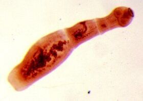 Infection occurs with food, coming into contact with animals and environmental objects inoculated with Echinococcus eggs. Once in the intestines, oncospheres (six-hooked larvae) develop from them. They enter the bloodstream from the intestines and can be carried throughout the body.
The worm's "favorite" parasite sites are the liver and lungs. Once settled in these organs, the larva becomes a Finn (echinococcus cyst), which gradually grows and begins to destroy nearby tissues. Often, echinococcosis is mistaken for benign or malignant tumors in the diagnostic process. In addition to the mechanical effect (compression of organs and blood vessels), cystic rupture of the echinococcus sometimes occurs. This condition can cause toxic shock or the formation of several new cysts.
Alveococcus
This parasite, considered a type of echinococcus, causes one of the most dangerous helminthiases (alveococcosis), similar in severity to cirrhosis and liver cancer. Infection occurs when oncospheres (eggs with mature larvae) enter the intestines. There, the embryo leaves the ovum and, penetrating the intestinal walls, enters the bloodstream. Furthermore, through blood flow, the parasite spreads to all tissues and organs in the body (most often localized in the liver). This is where the main developmental stage in the larvae begins (multi-chamber bubble, laurocyst is formed). In each chamber is the embryonic head of the parasite, which gradually develops further. Laurocysts are very aggressive formations that grow continuously due to growing bubbles and are also able to grow into the liver as metastases from cancer. Necrotic changes due to vascular dysfunction go through necrotic changes in nearby tissues. Spreading to nearby structures, the alveococcus forms fibrous nodules with inclusions of multi-chamber bubbles. This condition can last for several years and therefore requires mandatory surgery.
Diagnosis of helminthiasis
Diagnosis of helminthic invasions includes the following activities:
- Thorough medical history to help find possible causes of infection; Laboratory examination of
- feces, blood, 12p intestinal contents, rectal and perianal mucus, muscle tissue, lung sputum, bile. The analysis may reveal eggs, segments or fragments of the parasites. However, increased eosinophil levels in the blood also indicate the presence of helminthiasis.
- Serological tests (ELISA, RSK, indirect agglutination reaction, immunofluorescence analysis, etc. ) are performed when diagnosing diseases caused by larval stages or tissue parasites.
- Ultrasound, CT, and endoscopic examinations are prescribed to detect helminth samples affecting liver tissue.
Human worms: treatment
In the acute phase of a parasitic infection, the patient is prescribed detoxification and desensitization therapy. In severe cases of the disease (liver trematodes, trichinosis) glucocorticoids are used according to medical indications.
Special anthelmintic chemotherapeutic agents are prescribed as a specific therapy drug, taking into account the nature of the pathogen.
At the same time, the patient should take antihistamines and enterosorbents. The final stage of treatment involves the use of probiotics that normalize the intestinal microflora.
A special gentle diet is also prescribed (food must be digestible and low in fat).
During anthelmintic therapy, the patient should adhere to strict personal hygiene (to avoid re-infection). However, for many helminthiasis, all family members and individuals who are in constant contact with those infected should be treated.
Prevention of helminthiasis
- Maintaining personal and public hygiene;
- Strict adherence to cooking technology;
- Regular examination and preventive treatment of pets;
- Thorough washing of fresh vegetables, fruits and herbs;
- Proper management of river fish;
- Avoid eating raw, lightly salted and dried fish.